Cryo-EM: A Look Inside the Living Cell
By Nataliia Bohdanova
Introduction
Understanding what goes on inside a living cell has always fascinated scientists. But how can we actually see the tiny structures inside something so small? One powerful tool is cryo-electron microscopy, or cryo-EM. This technique freezes biological samples and uses a beam of electrons to take incredibly detailed images. Then, these images are combined to build a full 3D model of the cell’s inner machinery.
What Is Cryo-Electron Microscopy?
Cryo-EM is a form of electron microscopy where samples are rapidly frozen to preserve their natural state. Unlike traditional methods, it doesn’t require staining or slicing the sample thin. Freezing prevents the formation of ice crystals that could damage the cell, and helps keep delicate structures intact.
How It Works
- Freezing the Sample: A tiny drop of the biological material is placed on a grid and plunged into liquid ethane, freezing it almost instantly.
- Taking Electron Images: The frozen sample is placed in a microscope that uses electrons instead of light. Electron beams can reveal much smaller structures than light microscopes can.
- Image Collection: The microscope captures thousands of 2D images from different angles.
- 3D Reconstruction: Using computer algorithms, the images are combined to create a highly detailed 3D model.
Why It Matters
Cryo-EM allows scientists to see the shape of proteins, viruses, and cellular structures as they are in real life. It has been key to breakthroughs in medicine and biology, such as understanding how the coronavirus spike protein works.
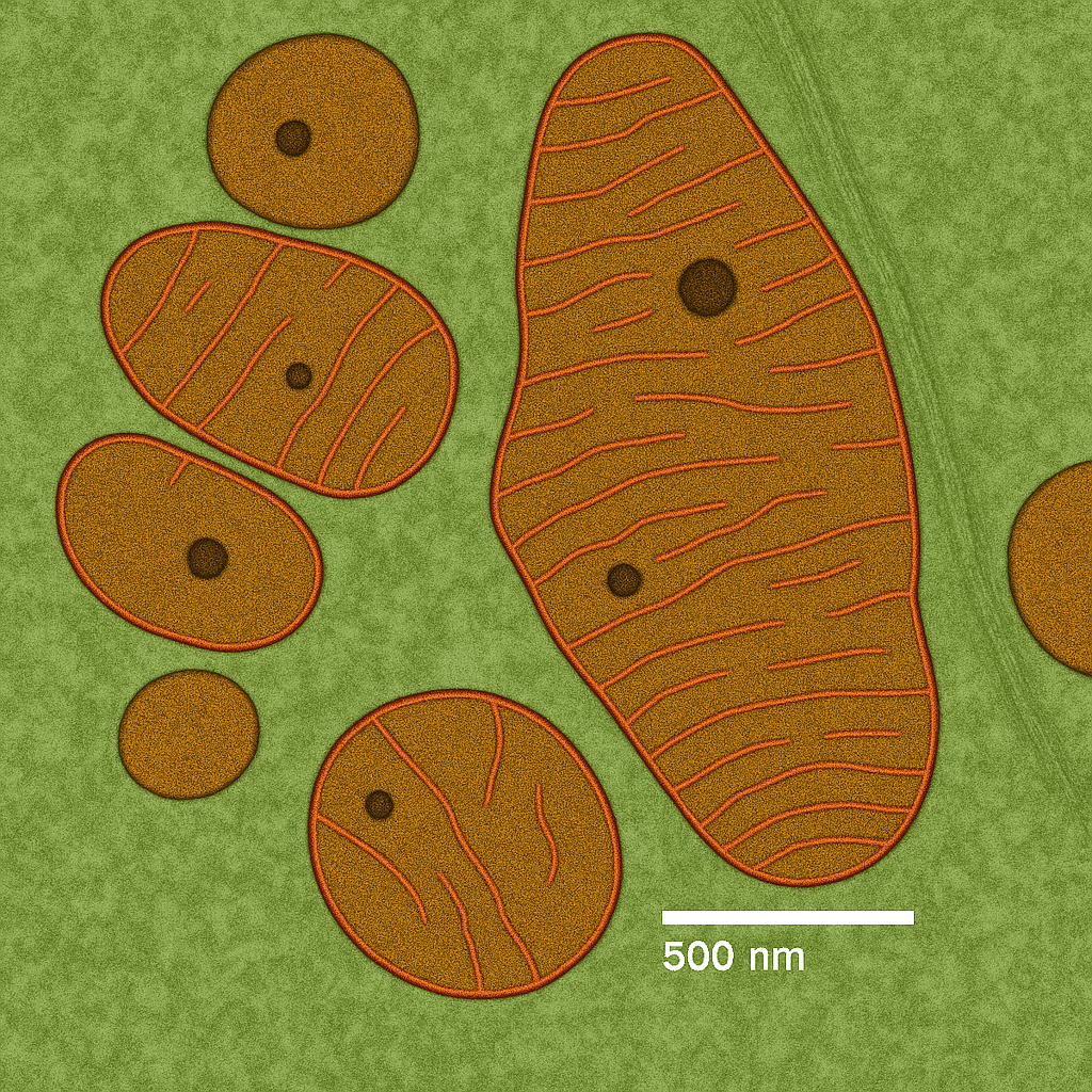
Image based on original photo from jonlieffmd.com, modified for illustration purposes.
Cryo-EM and Cells
One of the most exciting uses of cryo-EM is to model the interior of entire cells. By taking multiple “slices” and stitching them together, researchers can build a virtual 3D tour of a cell. This reveals organelles, protein complexes, and other inner components in unprecedented detail.

Source: Wellcome Images, CC BY 4.0
The Future of Cell Imaging
Cryo-EM is rapidly improving. With better detectors, software, and automation, it’s becoming faster and more accessible. In the future, scientists hope to watch processes inside cells almost in real time — a dream once considered science fiction.
Conclusion
Thanks to cryo-electron microscopy, we are now able to look inside the building blocks of life like never before. It’s a window into the microscopic world, helping us understand life from the inside out.
🧬 AI-Designed DNA Controls Gene Expression in Healthy Mammalian Cells | CRG Breakthrough 2025