


 In addition to organisms that have a cellular structure, there are non-cellular life forms – viruses and bacteriophages. These life forms are like a transitional group between animate and inanimate nature.
Viruses, in order to cope with the midst of the midst of the crystalline form, are called viruses. Virusi buli vidkriti in 1892 as a Russian student of D.I. Ivanovskiy. The Ukrainian word “virus” means “spit”.
Viruses that are in a crystalline form in the environment are called virions.
The viruses were discovered in 1892 p. Russian scientist DI Ivanovsky. Translated into Ukrainian, “virus” means “poison”.
Viruses are microscopic particles that exist almost everywhere on Earth. They are present in animals, plants, and other living organisms, and they can sometimes cause diseases.
The origin of viruses
Viruses have been found wherever there is life, and viruses have probably existed since the first living cells appeared. The origin of viruses is unclear, as they do not leave any fossils and their family ties can only be studied by molecular phylogenetics.
There are three main hypotheses about the origin of viruses:
• regression hypothesis;
• hypothesis of cellular origin (“mad genes” according to Watson)
• hypothesis of coevolution.
Regressive hypothesis
According to this hypothesis, viruses were once small eukaryotic cells parasitizing larger cells. Over time, these cells probably lost genes that were “superfluous” in the parasitic lifestyle. This hypothesis is based on the observation that some bacteria, namely rickettsiae and chlamydia, are cellular organisms that, however, like viruses, can only multiply inside another cell. This hypothesis is also called the degeneration hypothesis or the reduction hypothesis.
Bacteria → mycoplasmas and rickettsiae → chlamydia → smallpox viruses
Hypothesis of cellular origin, or the hypothesis of “mad genes”
Some viruses may have originated from DNA or RNA fragments “released” from the genome of a larger organism. Such fragments can be derived from plasmids (DNA molecules capable of being transferred from cell to cell) or from transposons (DNA molecules that replicate and move from place to place within the genome). Transposons, formerly known as “jumping genes”, are examples of mobile genetic elements, possibly from which some viruses may have originated. The transposons were discovered by Barbara McClintock in 1950 in corn. This hypothesis is also called the nomadic hypothesis or the escape hypothesis, the “mad gene hypothesis”, according to Watson.
Hypothesis of coevolution of viruses from precellular life forms
Hypothesis of coevolution of viruses from precellular life forms This hypothesis suggests that viruses originated in complex complexes of proteins and nucleic acids at the same time as the first living cells on Earth, and have depended on cell life for billions of years. In addition to viruses, there are other non-cellular life forms.
However, the hypothesis of “mad genes” does not explain the appearance of the capsid and other components of the viral particle. The hypothesis of coevolution contradicts the definition of viruses as non-cellular particles dependent on host cells.
Structure of viruses
Viruses are stored in DNA or RNA molecules, covered by a protein membrane, and some times a lipid membrane. Viruses can living in look like crystals (virioni). “Сrystalline virus” does not multiply, does not appear as a sign of the living, and it can be a long time. If a virus gets into cell, the virus will start to multiply, and all the structures of the cell will start to work for him. Penetrating into the cell, the virus inserts its nucleic acid into the DNA of the cell, the synthesis of viral particles proteins started, whereas the synthesis of cell proteins stops.
If the genetic apparatus of the virus is RNA, then first there is a process of reverse transcription according to the scheme RNA → DNA → RNA → protein, therefore RNA-dependent viruses are called retroviruses.
SHELL
Some viruses (complex) surround themselves with an additional (in addition to the protein capsid) shell of a modified cell membrane (plasma or internal, such as the nuclear membrane or the membrane of the endoplasmic reticulum). This additional bilipid layer is called the supercapsid.
The lipid envelope of the virus is permeated with proteins encoded by the viral genome and the genome of the host; the membrane itself, as well as any of its carbohydrate components originate entirely from the host cell. In this way, the influenza virus and HIV form their shell. The pathogenicity of most viruses depends on the presence of this shell.
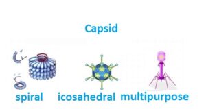
Thus, all viruses are divided into simple and complex. Simple viruses consist of a nucleic acid and a protein shell – the capsid; some crystallize; shape rod-shaped, filamentous and spherical. Complex viruses in addition to capsid proteins and nucleic acid may contain a lipoprotein membrane, carbohydrates and non-structural proteins – enzymes.
Life cycle
Viruses do not multiply by cell division, since they do not have a cellular structure. Instead, they use the resources of the host cell to form multiple copies of themselves, and their addition occurs within the cell.
Conventionally, the life cycle of a virus can be divided into several mutually overlapping stages (usually 6 stages are distinguished):
1. Attachment is the formation of a specific bond between viral capsid proteins and receptors on the surface of the host cell. This specific binding determines the host range of the virus. For example, HIV infects only a specific type of human leukocyte called helper T cells.
2. Penetration into the cell. At the next stage of the virus, it is necessary to deliver its own genetic material into the cells. Some viruses also transfer their own proteins into the cell, which are necessary for its implementation (this is especially true for viruses containing negative RNA).
3. Detachment of membranes is the process of loss of the capsid. This is achieved using viral enzymes or host cell enzymes. Ultimately, the viral genomic nucleic acid is released.
4. Replication of viruses involves, first of all, the replication of the genome. Virus replication includes the synthesis of mRNA of early viral genes (with the exception of viruses containing positive RNA), the synthesis of viral proteins, in a word, the virus is diluted with the help of the protein-synthesizing apparatus of the cell.
5. Following this, self-assembly of viral particles occurs, later some modifications of proteins occur. In viruses such as HIV, this modification (sometimes called maturation) occurs after the virus leaves the host cell.
6. Exit from the cell. Viruses can leave a cell after lysis, a process in which a cell dies due to rupture of the membrane and cell wall, if any. This feature is found in many bacterial and some animal viruses. Some viruses undergo a lysogenic cycle, where the viral genome is incorporated by genetic recombination into a special place on the host cell’s chromosome. Then the viral genome is called a provirus, or, in the case of a bacteriophage, a prophage.
When a cell divides, the viral genome also doubles. Within the cell, the virus generally does not manifest itself; however, at some point, the provirus or prophage can cause the activation of the virus, which can cause lysis of the host cells.
Even when the virus is actively multiplying, it does not always kill the host cell. Enveloped viruses, including HIV, are usually separated from the cell by freezing. During this process, the virus acquires its own envelope, which is a modified fragment of the host’s cell membrane.
In tests, the following question is encountered: “How do viruses multiply?” – to which we answer – “by self-assembly”. Viruses perform this function in the middle of the host cell, after the latter has reproduced the proteins of the virus.
BACTERIOPHAGES
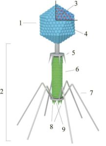 1-head, 2-process, 3-nucleic acid, 4-capsid, 5-collar, 6-sheath, 7-tail filaments, 8-spines, 9-basal plate
Bacteriophages are viruses that infect bacterial cells. The body of a bacteriophage consists of a protein head, in the center of which is the viral DNA, and a tail. At the end of the tail there are tail processes, which serve to fix them on the surface of the bacterial cell, and an enzyme that dissolves the membrane of the bacterial cell. Through the channel located in the tail, the virus introduces DNA into the bacterial cell and inhibits the synthesis of bacterial proteins, instead of which DNA and virus proteins are synthesized. In the cell, new viruses are formed, which leave dead bacteria and penetrate into new ones. Bacteriophages can be used as a medicine against pathogens of infectious diseases (cholera, typhoid fever).
According to the method of interaction with the host cell, the following types of infection are distinguished
- Lytic pathway – the virus attaches to the host cell and injects its nucleic acid into it, forcing the cell to produce daughter viral particles (progenetic or secondary, viruses) in huge quantities. Ultimately, this process leads to cell lysis – it bursts and a new generation of virions is released, ready to infect other cells.
- Lysogenic pathway (persistent, stable) – the virus in the cell is incorporated into the host’s DNA. Viral particles leave the cell gradually. The cell continues to live and divide, although its functioning may change. When cell DNA replicates, the viral DNA replicates at the same time. As a result of cell division, new copies of viral DNA are formed, embedded in the cell genome. Therefore, lysogeny is characterized by the insertion of a viral nucleic acid into the chromosome of a host cell in such a way that there is a potential for the transfer of the obtained genetic material to daughter cells during cell division. In this state, the information contained in the viral nucleic acid is not expressed.
- Latency is a phenomenon when a cell, in which a virus reproduces autonomously, retains its viability for a long time, is called latency. An actively multiplying virus does not always kill the host cell. Enveloped viruses, including HIV, are usually separated from the cell by freezing. During this process, the virus takes on a shell, which is a modified fragment of the host’s cell membrane or other inner membrane. In this way, the cell can continue to live and produce the virus. Viruses with such an infection are not released into the environment. Only certain factors can trigger the activation of the virus. Herpes is activated by a drop in immunity in case of colds.
VIRUSES CAUSING DISEASES
Poliomyelitis. It is mostly asymptomatic or erased. But sometimes the poliovirus enters the CNS and multiplies in motoneurons, which leads to their death, irreversible paresis or paralysis of the muscles innervated by them.
Measles is an acute infectious viral disease with a very high level of infectiousness, the causative agent of which is the measles virus. It is characterized by high fever (up to 40.5 ° C), inflammation of the mucous membranes of the oral cavity and upper respiratory tract, conjunctivitis and a characteristic maculopapular rash of the skin, general intoxication.
Rubella, or the third disease, is an epidemic viral disease with an incubation period of about 15-24 days. It is usually a harmless condition that mostly affects children, but it can cause serious birth defects if a woman becomes infected early in pregnancy. The name “third disease” comes from the times when a list of diseases provoking childhood rashes was compiled, in which rubella was in third place.
Сhickenpox (lat. Varicella) is an acute highly contagious viral disease with airborne transmission. Usually characterized by a febrile condition, papulovesicular rash with a benign course. The causative agent of chickenpox is the varicella-zoster virus, lat. Varicella Zoster of the Herpesviridae family, also called the third type of herpesvirus (Human herpesvirus 3). Along with chickenpox, it is the causative agent of herpes zoster (shingles).
Rabies (rabies) is a natural focal especially dangerous deadly infectious disease caused by the rabies virus. Rabies belongs to the group of so-called neglected diseases due to its low prevalence and incidence in developed countries. Passed with saliva when bitten by sick animals. Then, spreading along the nerve pathways, the virus reaches the salivary glands, nerve cells of the cerebral cortex, hippocampus, bulbar centers and, affecting them, causes severe disorders leading to death.
Parotitis is an inflammation of the parotid salivary gland. The name comes from ancient Greek. παρα- – near, near and οὖς, genitive ὠτός – ear. Mumps is specific and nonspecific. There are both infectious and autoimmune and non-infectious parotitis, the cause of the latter may be dehydration, trauma or prolonged hypothermia followed by inflammation of the parotid salivary glands.
Hepatitis (Greek ἡπατῖτις, from ἥπαρ – liver) – inflammatory liver diseases of various, including viral etiology. In a general sense, it characterizes any inflammation of the liver. In 2016, a large international study found that deaths from hepatitis are comparable to those of tuberculosis, malaria and HIV. Human Immunodeficiency Virus (HIV) is a retrovirus from the genus of lentiviruses that causes a slowly progressive disease – HIV infection. The virus infects cells of the immune system that have CD4 receptors on their surface: T-helpers, monocytes, macrophages, Langerhans cells, dendritic cells, microglia cells. As a result, the immune system is suppressed and acquired immune deficiency syndrome (AIDS) develops.
INFECTIOUS AGENTS
Prions are isoforms of common proteins of the nervous system, differing in the way they fold the polypeptide chain. They affect the central nervous system of vertebrates using the energy reserves of neurons.
For a long time, doctors could not identify the causes of Cranefe Ice-Jacobs disease, Gerstmann-Strausler-Schenkler syndrome, multiple sclerosis, Vilyui encephalitis, chicken, leukospongiosis, fatal familial insomnia, etc. In animals, slow infections include skreley, transmissible spongiform encephalopathy, mink encelopathy, debilitating disease of deer, moose, mules, etc. All these diseases occur with severe lesions of the central nervous system and develop over the years.
The nature of “slow infections” was established in the 80s of the XX century. The studies of the American scientist D. Gaidushek revealed that slow viruses are a fundamentally new type of pathogenic agent – an infectious protein. Prions were identified by the American researcher S. Prusiner. It turned out that these are proteins with a molecular weight of 35–105 kDa and a length of 50–150 nm. Prions, similar to proteins in the brain, have the ability to penetrate the body and damage the central nervous system, causing gradual degradation of neurons. The incubation period for prion infections is 3-9 months to 2-5 years. Prion infections were first talked about at the turn of the millennium. This is due to the epidemic of mad cow disease (transmissible spongiform encephalopathy), which has hit livestock farms in Europe. This disease also affects humans, which makes it especially dangerous.
Viroids are small circular RNA molecules that do not encode any proteins. They parasitize higher plants and reproduce only with the help of plant cell enzymes.
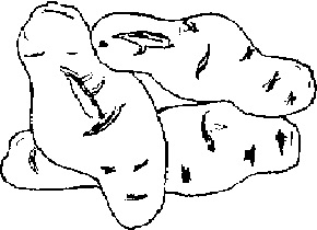 spindle-like potato tubers caused by viroids
Viroids were discovered in the mid-70s. XX century This is a circular RNA 300-400 nucleotides long. About 30 faiths are known. They all turned out to be plant parasites. Viroids lack a protein capsid, which makes them unable to enter intact cells. They pass from plant to plant only when both the donor cell and the recipient cell are damaged.
So:
Viruses are infectious particles composed of nucleic acid molecules packed in a protein coat (capsid). They act as parasites of most prokaryotic and eukaryotic cells.
Viroids are small circular RNA molecules that do not encode any proteins. They parasitize higher plants and reproduce only with the help of plant cell enzymes.
Prions are isoforms of common proteins of the nervous system, differing in the way they fold the polypeptide chain. They affect the central nervous system of vertebrates using the energy reserves of neurons.
Antiviral drug


